If your OB or midwife has recommended a fetal echocardiogram, it is natural to wonder what they are looking for. For most families, this test is a careful, extra look at the baby’s heart, and most results come back normal. Knowing why it was ordered and what happens during the visit can take a lot of the worry out of the appointment.
What Is a Fetal Echocardiogram?
A fetal echocardiogram is a detailed ultrasound focused entirely on your baby’s heart. It looks at the heart’s four chambers, the valves, the major blood vessels, and the heart’s rhythm. It also shows how blood flows through each structure.
Your standard anatomy scan includes a quick check of the heart. A fetal echo goes much deeper, using specialized views and more time to evaluate details a routine scan is not designed to catch.
Why Heart Screening Matters
Congenital heart defects are the most common type of birth defect. According to the CDC, they affect about 1 in every 100 babies born in the United States each year. Some are minor and never need treatment. Others require care right after birth.
Finding a heart condition before delivery allows your team to plan ahead. That can mean delivering at a hospital with a pediatric cardiology team ready, rather than arranging a transfer after the baby arrives.
Who Needs a Fetal Echocardiogram?
Most fetal echo referrals come down to a handful of common reasons. Having one of these factors does not mean something is wrong with your baby’s heart. It means a closer look is worthwhile.
| Reason for Referral | Common Examples |
|---|---|
| Family history | A parent or sibling with a congenital heart defect |
| Parent’s health | Diabetes before pregnancy, certain autoimmune antibodies, some seizure or acne medications |
| How the pregnancy began | Conception through IVF |
| Findings during pregnancy | An unclear heart view on the anatomy scan, an irregular heartbeat, extra fluid around the baby |
| Screening or genetic results | Increased neck fluid on first trimester screening or a chromosomal finding |
| Multiple pregnancy | Identical twins who share a placenta |
If you have lupus or Sjögren’s antibodies, our post on lupus and pregnancy explains why heart rhythm monitoring may be part of your care.
Has your provider recommended a fetal echocardiogram? Schedule a consultation with Lancaster Maternal Fetal Medicine and get an expert look at your baby’s heart, close to home.
When Is a Fetal Echo Done?
Most fetal echocardiograms take place between about 18 and 24 weeks of pregnancy. By then, the heart is large enough to see clearly, and there is still time to plan if anything needs follow-up. In some cases, an early look happens sooner, with a repeat scan later in pregnancy.
What to Expect During the Appointment
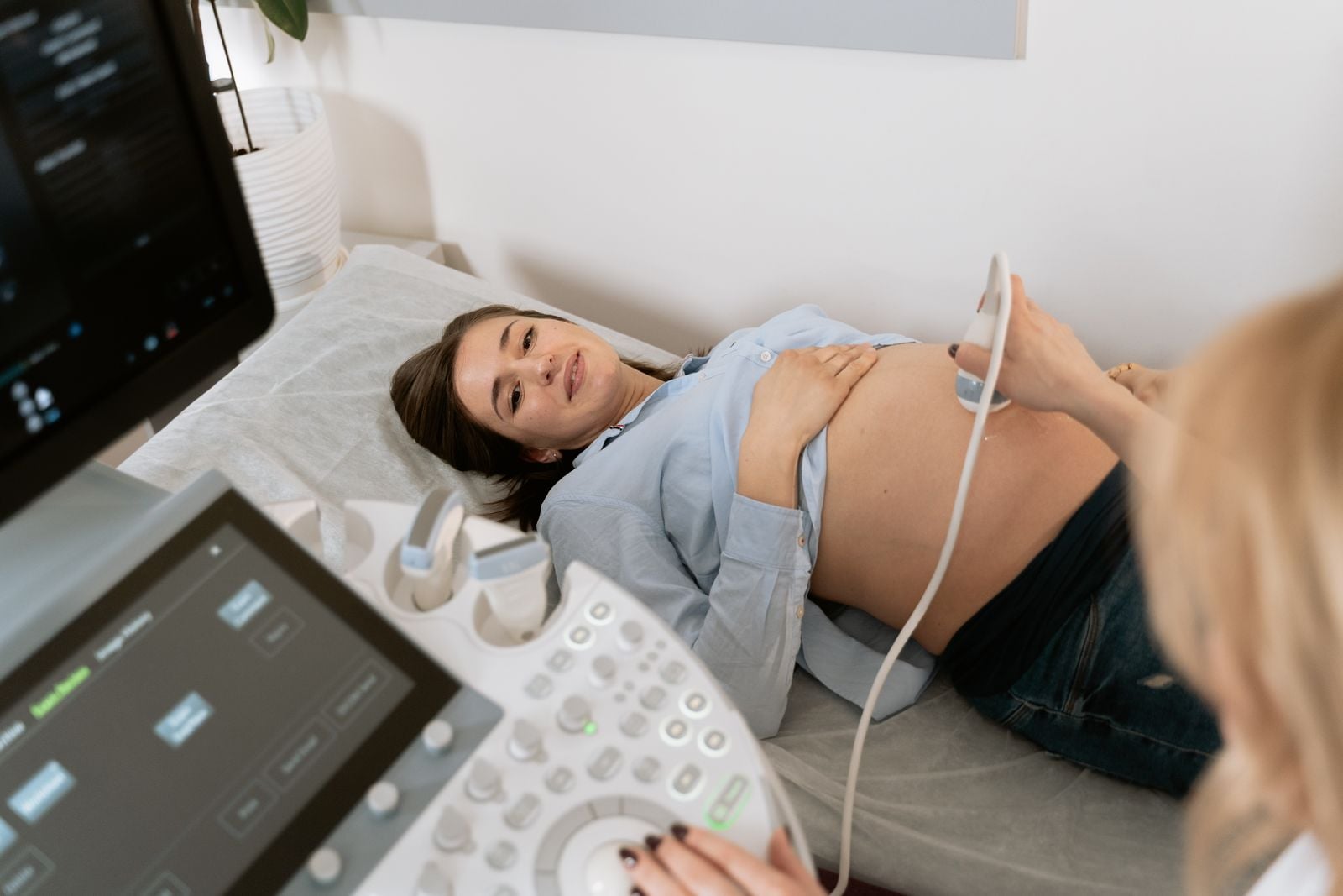
A fetal echo feels much like any other pregnancy ultrasound. You lie back comfortably while a specialist moves a probe with warm gel across your belly. There are no needles and no radiation.
The scan usually takes 30 to 60 minutes. It can take a little longer if your baby is in a tricky position, so a short walk or a change of position may help. Afterward, a specialist reviews the images and talks through the results with you.
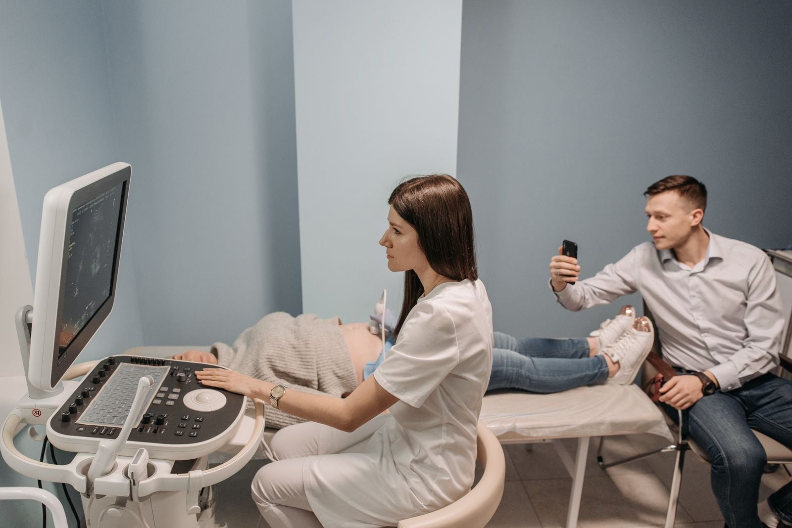
It is completely normal to feel nervous during the scan, especially if the room is quiet while the specialist concentrates. Silence usually just means careful work. Feel free to ask questions as you go, and bring a partner or support person if that helps you feel more at ease. Many parents find it reassuring to see the heart beating on screen and hear each part explained.
What Happens If Something Is Found?
If a heart condition is identified, you will not be left to figure out next steps alone. Your team will explain the finding in plain language, discuss any follow-up imaging, and coordinate with pediatric cardiology specialists. Together, you will build a delivery plan so your baby has the right care from the very first moments. Many babies with heart conditions grow up to live full, active lives, and early planning is a big part of that.
Fetal Echocardiography in Lancaster County
Families across Lancaster County, the Belleville area, and the greater Reading region rely on Lancaster Maternal Fetal Medicine for fetal echocardiography because our doctors and sonographers are specially trained in prenatal heart imaging. We also work with nationally accredited pediatric cardiology centers, so if your baby needs more specialized care, the handoff is smooth.
We partner with your OB or midwife, sending results back quickly so everyone stays on the same page.
Frequently Asked Questions
Is a fetal echocardiogram safe for my baby?
Yes. A fetal echocardiogram uses the same sound-wave technology as a standard pregnancy ultrasound. It involves no radiation and no needles, and it is safe for both you and your baby.
How long does a fetal echocardiogram take?
Most fetal echocardiograms take 30 to 60 minutes. The scan can run longer if your baby’s position makes certain heart views hard to capture.
Do I need to prepare for a fetal echocardiogram?
Usually no special preparation is needed. Wear comfortable, two-piece clothing so your belly is easy to reach, and follow any instructions our office gives you when scheduling.
Can a fetal echocardiogram detect every heart problem?
A fetal echocardiogram detects most major structural heart defects, but no test is perfect. Some small defects, and certain conditions that only appear after birth, may not be visible before delivery.
What happens if my fetal echocardiogram is normal?
If the results are normal, you will typically continue your regular prenatal care. Your specialist may still recommend a follow-up scan if there are ongoing risk factors, such as diabetes or certain antibodies.