
Pioneering 3D Ultrasound Imaging
By Dr. Christian Macedonia on March 16, 2022

Dr. Macedonia was the first person to produce a 3D ultrasound image of a living fetus.
The day the world’s first 3D ultrasound of a living fetus was taken was an ordinary winter day in Bethesda, Maryland.
It was cold and windy on December 19, 1991. I was a medical student at Uniformed Services University, working as a researcher at the National Institutes of Health across the street. The medical school had allowed me to do an independent study project and I had chosen “3D Ultrasound.” Most people thought I was joking. Fortunately for me, a few computer scientists at NIH took me seriously.
While there had been a few 3D ultrasounds performed on fetuses up to that point, none had been done on a living fetus. It was easier for researchers to take multiple still shots of a non-moving fetus, so work had been done exclusively on stillborn babies, either still in the womb or placed into water tanks.
Conventional ultrasound takes 2D pictures and updates the images typically between 20-50 times a second. That was the state of the technology in 1991. I figured that if 2D ultrasound takes “slices” then I could simply run the probe along a track in a third dimension and capture all the frames to make a stack of images. I could then render the stack in 3D. It is easy to think about if you consider this to be like a loaf of bread. Each image is a slice. Put all the slices together and you have a loaf.
For me to make this vision of the future happen I needed a few things: I needed access to an ultrasound machine, a volunteer mom, a machine to make the ultrasound probe move in a precise manner in a straight line, and rendering software to put the stack together and render in 3D.
Putting the machine together was easy for me. I had some basic training in electronics and physics and circuits from my undergrad days at Bucknell University. It took me about a week to make the first probe, working in my basement workshop, using supplies from Hechinger’s Hardware that included a cordless screwdriver, a threaded dowel, and a lab clamp.
I was lucky enough to have met Dr. Wayne Rasband at the NIH who had developed a piece of software called NIH Image (now called “Image J”). It was a general purpose image processing toolkit, used mostly by NIH researchers to capture microscope slide images. It had never been used for ultrasound before. A few simple modifications had to be made to get it to work with ultrasound images I collected on VHS.
My wife and I were expecting our first child. She herself was a researcher at the NIH, working on cures for infectious diseases. The chief of radiology at the NIH gave us access to his best 2D ultrasound machine available, the Accuson 128, in the basement of Building 12. Our baby, (who is now 29 years old!), was 20 weeks along at the time.
On that cold December day, we tried the system out. I took the tapes of the sweeps to Dr. Rasband who ran the captures of the slices into NIH Image. After 90 minutes of calculation, the world’s first 3D ultrasound of a living fetus appeared on the screen.
The first time the public saw the images of that first 3D ultrasound was at the 37th Annual American Institute of Ultrasound Annual Scientific Meeting in Honolulu, Hawaii on the 15th of March, 1993. It was displayed as part of the opening session of the convention. I have been to many scientific meetings in my lifetime, but this one is the most memorable for me. When I hit “play” on my computer and showed the 3D ultrasound rotating on the screen, there was an audible gasp of amazement from the thousand delegates in the audience. Awards and accolades soon followed, along with a $5 million scientific grant to continue the work.
Of course, I worked for the government (in fact, I was a second lieutenant in the US Army at the time) so there was no profit or patent for me. What it did allow me to do was follow my dreams, explore, and have the quiet satisfaction that our family had not only given birth to our precious newborn boy, but also a scientific revolution that has literally changed the way our society views the world of the unborn.
Here are a few modern 3D ultrasound images, taken at Lancaster MFM:

First trimester fetus
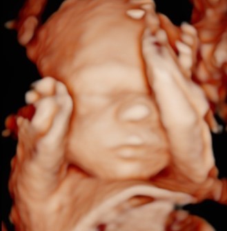
Third trimester fetus
For more information on ultrasound during pregnancy visit our blog post, “Ultrasound: How, When and Why” at https://lancastermfm.com/ultrasound-how-when-and-why/
*Lancaster MFM does not recommend the use of “keepsake” 3D ultrasound companies. Ultrasound is safe, but should only be used by a qualified provider, and as little as possible. As you can see from the images above, our obstetric sonographers are VERY good at getting nice 3D images!
Here’s a summary of the FDA guidance, put out in 2014: